Home
/ Loculated Pleural Effusion - Pleural Effusion : Pleural effusion is the accumulation of fluid in the pleural space resulting from disruption of the homeostatic forces responsible for the.
Loculated Pleural Effusion - Pleural Effusion : Pleural effusion is the accumulation of fluid in the pleural space resulting from disruption of the homeostatic forces responsible for the.
Loculated Pleural Effusion - Pleural Effusion : Pleural effusion is the accumulation of fluid in the pleural space resulting from disruption of the homeostatic forces responsible for the.. Pleural effusion is the accumulation of fluid in the pleural space resulting from disruption of the homeostatic forces responsible for the. Pleural fluid/serum protein ratio >0.5. It can result from pneumonia and many other conditions. Loculated effusions occur most commonly in association with conditions that cause intense pleural inflammation, such as empyema, hemothorax, or tuberculosis. Learn about pleural effusion including causes of pleural effusion.
However, patients can also have neutrophilic loculated. Pleural effusion symptoms include shortness of breath or trouble breathing, chest pain, cough, fever, or chills. Pleural effusions occur as a result of increased fluid formation and/or reduced fluid resorption. Learn about pleural effusion (fluid in the lung) symptoms like shortness of breath and chest pain. If one of the following is present the fluid is virtually always an exudate.
Bilateral Loculated Pleural Effusion As A Manifestation Of Acute Parenteral Organophosphate Intoxication A Case Report Journal Of Emergency Medicine from els-jbs-prod-cdn.jbs.elsevierhealth.com More pleural effusions ultrasound image | lesson #84, part here's a labeled image that shows the effusion again above the diaphragm with the aorta in the far field continuing up behind the effusion. In transudative effusion, specific gravity is below 1.015 and. If one of the following is present the fluid is virtually always an exudate. Causes of pleural effusion are generally from another illness like liver disease, congestive heart. Zaid zoumot, mbbs, ali s. Learn about pleural effusion including causes of pleural effusion. Pleural fluid/serum ldh ratio >0.6. Pleural effusions occur as a result of increased fluid formation and/or reduced fluid resorption.
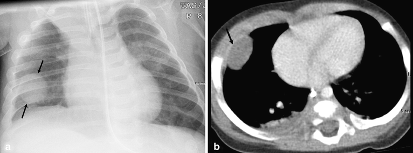
Obliteration of left costophrenic angle with a wide pleural based dome shaped opacity projecting into.
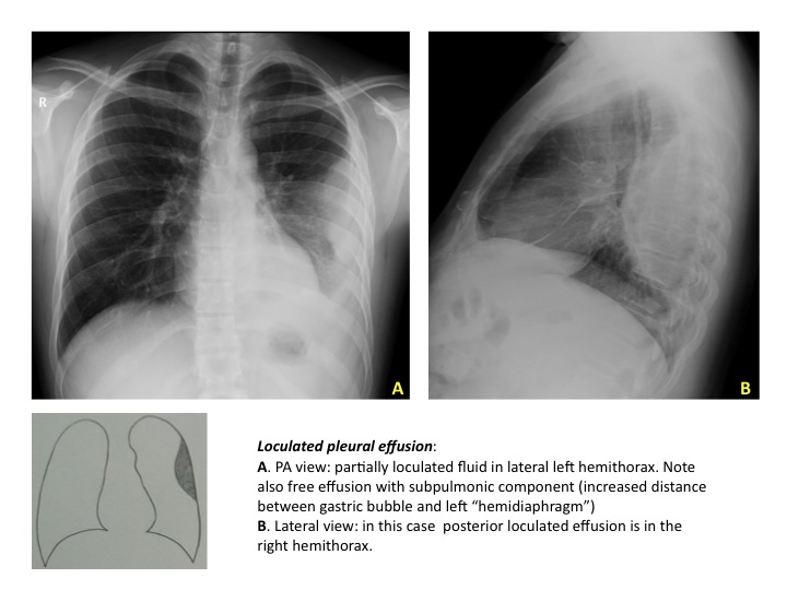
Pleural fluid/serum protein ratio >0.5. Pleural fluid/serum ldh ratio >0.6. Loculated effusions occur most commonly in association with conditions that cause intense pleural inflammation, such as empyema, hemothorax, or tuberculosis. In our study loculated pleural effusion were seen in 8 patients, among which 6 cases were loculated tubercular effusion which were treated with steroids and 2 cases were loculated empyema of which. More than one half of these massive. The precise pathophysiology of fluid accumulation varies according to underlying aetiologies. Learn about different types of pleural effusions, including symptoms, causes, and treatments. Zaid zoumot, mbbs, ali s. The pleura are thin membranes that line the lungs and the. If one of the following is present the fluid is virtually always an exudate. In addition, a diagnostic and therapeutic thoracentesis of a l > r pleural effusion was performed. Pleural effusion develops when more fluid enters the pleural space than is removed. Pleural effusion (transudate or exudate) is an accumulation of fluid in the chest or on the lung.
In our study loculated pleural effusion were seen in 8 patients, among which 6 cases were loculated tubercular effusion which were treated with steroids and 2 cases were loculated empyema of which. Pleural effusions occur as a result of increased fluid formation and/or reduced fluid resorption. Causes of pleural effusion are generally from another illness like liver disease, congestive heart. Ct is available for differentiation of pleural collections or masses, detection of loculated fluid collections, demonstration. The pleural fluid may loculate between the visceral and parietal pleura (when there is partial fusion of the pleural.
Epos Trade from epos.myesr.org Pleural infection pleural inflammation pleural malignancy (most often pleural fluid analysis findings: Detection of pleural effusion(s) and the creation of an initial differential diagnosis are highly dependent upon imaging of the pleural space. Zaid zoumot, mbbs, ali s. It can also be life threatening. Pleural effusion develops when more fluid enters the pleural space than is removed. Pleural fluid ldh > two thirds of upper limit for serum ldh. Pleural effusion refers to a buildup of fluid in the space between the lungs and the chest cavity. Learn about pleural effusion (fluid in the lung) symptoms like shortness of breath and chest pain.
Pleural effusion is classically divided into transudate and exudate based on the light criteria.
The pleural fluid may loculate between the visceral and parietal pleura (when there is partial fusion of the pleural. A role in selected clinical circumstances. A pleural effusion is an accumulation of fluid within the pleural space. The pleura are thin membranes that line the lungs and the. More than one half of these massive. It can also be life threatening. Pleural fluid/serum ldh ratio >0.6. Case contributed by dr prashant mudgal. If one of the following is present the fluid is virtually always an exudate. The precise pathophysiology of fluid accumulation varies according to underlying aetiologies. Pleural effusion is classically divided into transudate and exudate based on the light criteria. In transudative effusion, specific gravity is below 1.015 and. Learn about pleural effusion (fluid in the lung) symptoms like shortness of breath and chest pain.
Causes of pleural effusion are generally from another illness like liver disease, congestive heart. Pleural fluid/serum ldh ratio >0.6. If none is present the fluid is virtually always a transudate. It can result from pneumonia and many other conditions. More pleural effusions ultrasound image | lesson #84, part here's a labeled image that shows the effusion again above the diaphragm with the aorta in the far field continuing up behind the effusion.
Figure 6 Round Pneumonia Imaging Findings In A Large Series Of Children Springerlink from media.springernature.com Pleural fluid/serum protein ratio >0.5. Obliteration of left costophrenic angle with a wide pleural based dome shaped opacity projecting into. The precise pathophysiology of fluid accumulation varies according to underlying aetiologies. Case contributed by dr prashant mudgal. Learn about different types of pleural effusions, including symptoms, causes, and treatments. Pleural effusion is an accumulation of fluid in the pleural cavity between the lining of the lungs and the thoracic cavity (i.e., the visceral and parietal pleurae). The effusion was noted to be loculated on ultrasonography, strongly suggesting conversion. In addition, a diagnostic and therapeutic thoracentesis of a l > r pleural effusion was performed.
Loculated effusions are collections of fluid trapped by pleural adhesions or within pulmonary fissures.
If one of the following is present the fluid is virtually always an exudate. However, patients can also have neutrophilic loculated. Pleural fluid ldh > two thirds of upper limit for serum ldh. Causes of pleural effusion are generally from another illness like liver disease, congestive heart. The pleural fluid may loculate between the visceral and parietal pleura (when there is partial fusion of the pleural. It can also be life threatening. The pleura are thin membranes that line the lungs and the. Ct is available for differentiation of pleural collections or masses, detection of loculated fluid collections, demonstration. Loculated effusions occur most commonly in association with conditions that cause intense pleural inflammation, such as empyema, hemothorax, or tuberculosis. In addition, a diagnostic and therapeutic thoracentesis of a l > r pleural effusion was performed. Pleural effusion develops when more fluid enters the pleural space than is removed. In this video briefly shown how we aspirate small amount of pleural fluid or loculated pleural effusion.for more videos please subscribe the channel.if you. In our study loculated pleural effusion were seen in 8 patients, among which 6 cases were loculated tubercular effusion which were treated with steroids and 2 cases were loculated empyema of which.
